
Multisystem inflammatory syndrome in children (MIS-C).A chest X-ray usually is taken after placement of such medical devices to make sure everything is positioned correctly. Catheters are small tubes used to deliver medications or for dialysis. Pacemakers and defibrillators have wires attached to your heart to help control your heart rate and rhythm. A pacemaker, defibrillator or catheter.Your doctor can look at any lines or tubes that were placed during surgery to check for air leaks and areas of fluid or air buildup. Chest X-rays are useful for monitoring your recovery after you've had surgery in your chest, such as on your heart, lungs or esophagus. Rib or spine fractures or other problems with bone may be seen on a chest X-ray.

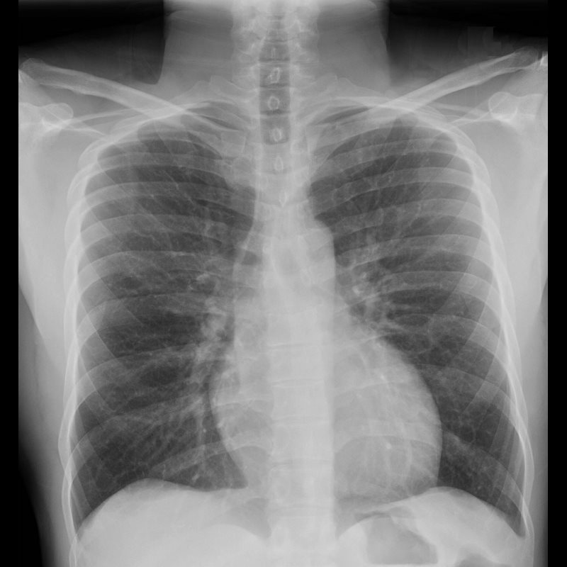
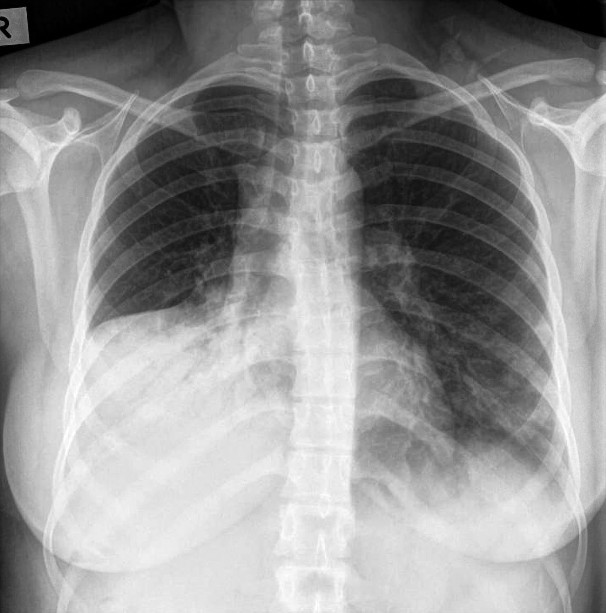
Calcified nodules in your lungs are most often from an old, resolved infection. Its presence may indicate fats and other substances in your vessels, damage to your heart valves, coronary arteries, heart muscle or the protective sac that surrounds the heart. Chest X-rays can detect the presence of calcium in your heart or blood vessels. Because the outlines of the large vessels near your heart - the aorta and pulmonary arteries and veins - are visible on X-rays, they may reveal aortic aneurysms, other blood vessel problems or congenital heart disease. Changes in the size and shape of your heart may indicate heart failure, fluid around the heart or heart valve problems. For instance, fluid in your lungs can be a result of congestive heart failure. Chest X-rays can show changes or problems in your lungs that stem from heart problems. They can also show chronic lung conditions, such as emphysema or cystic fibrosis, as well as complications related to these conditions. Chest X-rays can detect cancer, infection or air collecting in the space around a lung, which can cause the lung to collapse. A chest X-ray can also be used to check how you are responding to treatment.Ī chest X-ray can reveal many things inside your body, including: A chest X-ray is often among the first procedures you'll have if your doctor suspects heart or lung disease. Talk to your doctor if you are worried about the possible effects of x-rays.Chest X-rays are a common type of exam. The benefits of finding out what is wrong outweigh any risk there may be from radiation. The risk of the radiation causing any problems in the future is very small. The amount of radiation you receive from an x-ray is small and doesn't make you feel unwell. Your doctor and radiographer make sure the benefits of having the test outweigh these risks. Possible risksĪn x-ray is a safe test for most people but like all medical tests it has some possible risks. After your chest x-rayĪfter the x-ray you can get dressed and go home or back to work. So the whole process might take a few minutes. You usually need to have more than one x-ray taken from different angles. They might ask you to hold your breath for a few seconds. The radiographer then goes behind a screen to take the x-ray. The radiographer lines the machine up to make sure it is in the right place. The radiographer will help you to get into the correct position. If you can't stand you can have it sitting or lying on the x-ray couch. You usually have a chest x-ray standing up against the x-ray machine. Women may need to remove their bras as metal clips and underwiring can show up on the x-ray.


When you arrive in the x-ray department, the radiographer might ask you to change into a hospital gown. You can eat and drink normally beforehand. There is no special preparation for an x-ray. You have the x-ray in the x-ray or imaging department of the hospital. If you have symptoms that could be caused by lung cancer your doctor will arrange for you to have an x-ray. Changes can be due to cancer but can also be caused by other lung conditions. X-rays use high energy rays to take pictures of the inside of your body.


 0 kommentar(er)
